
News and Announcements
Acetabular labral tear: Etiology, Diagnostic Approach & Arthroscopic Treatment
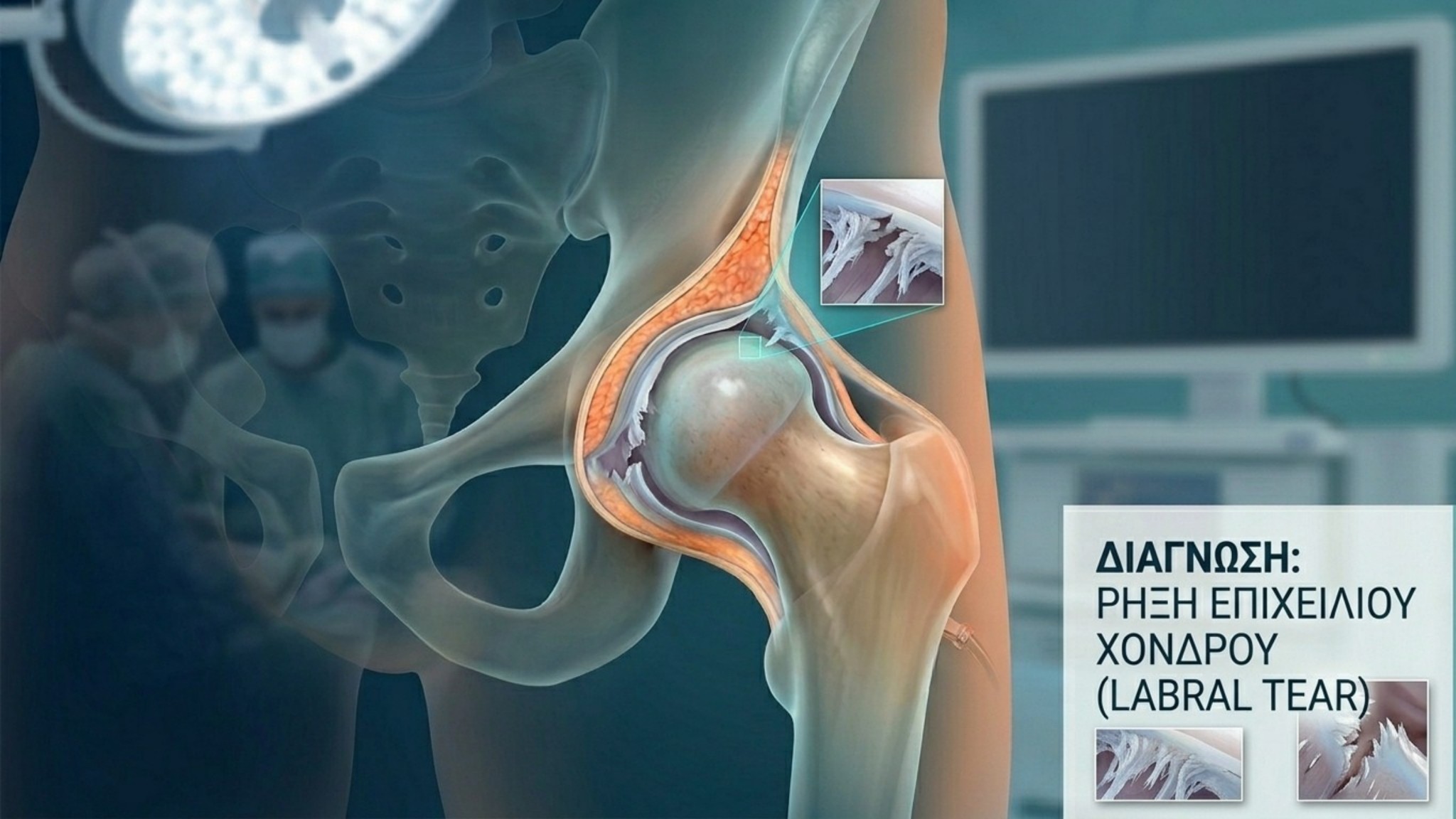
The acetabular labrum is a fibrocartilaginous ring surrounding the acetabulum, providing a mechanical "suction seal" and dynamic stability to the hip joint. Its structural integrity is critical for proper load distribution and protection of the articular cartilage. A labral tear represents one of the most common causes of anterior hip pain in young and active adults.
Clinical Presentation & Symptoms
Diagnosis relies on a thorough clinical evaluation of symptoms, which typically manifest gradually:
-
Anterior Hip Pain: Primarily localized in the groin area, characteristically presenting with the "C-sign," where the patient delineates pain by placing their hand over the greater trochanter.
-
Mechanical Symptoms: Sensations of clicking, locking, or catching throughout the range of motion.
-
Exacerbation in Sitting: Pronounced discomfort during prolonged hip flexion (e.g., driving, low seating).
-
Functional Limitation: Stiffness and reduced range of motion, particularly in flexion and internal rotation.
Etiological Factors
Labral tears are generally the result of underlying mechanical joint conditions or progressive wear:
-
Femoroacetabular Impingement (FAI): Osseous prominence on the femoral head/neck (Cam-type) or acetabulum (Pincer-type) leads to repetitive micro-trauma and progressive labral detachment.
-
Athletic Stress: Activities involving extreme rotational motions or hyperextension (soccer, dance, martial arts).
-
Acute Trauma: Sudden injuries, rotational trauma, or joint dislocations.
-
Degenerative Tear: Develops gradually from natural joint wear, chronic stress, or early osteoarthritis. The labrum loses elasticity, weakens structurally, and undergoes fibrillation or tearing without a discrete acute injury.
Diagnostic Protocol
An accurate diagnosis requires a combination of targeted imaging modalities:
-
Radiographic Evaluation: Specialized views (e.g., Dunn view) to assess bone morphology and rule out FAI or dysplasia.
-
Magnetic Resonance Imaging / Arthrography (MRI / MRA): The gold standard for visualizing the tear and evaluating the underlying articular cartilage quality.
Arthroscopic Reconstruction & One Day Clinic Protocol
When conservative management fails, Hip Arthroscopy offers a definitive, minimally invasive solution:
-
Anatomic Labral Repair: The detached labrum is refixed to the acetabular rim using bioabsorbable anchors, fully restoring the mechanical suction seal.
-
Correction of Underlying Deformity (Osteoplasty): Precise resection of the bony prominence (Cam/Pincer) eliminates impinging contact and prevents recurrence.
Under the specialized One Day Clinic protocol, the procedure is performed with minimal soft-tissue trauma. The patient mobilizes immediately on the day of surgery and begins a targeted rehabilitation program for a safe return to daily and athletic activities.
Frequently Asked Questions (FAQ)
1. Can an acetabular labral tear heal on its own? The acetabular labrum has a limited blood supply, particularly at its free margin. Consequently, a full tear or detachment cannot self-heal without surgical intervention. However, symptoms may temporarily subside with non-operative management.
2. What happens if a labral tear is left untreated? Loss of the suction seal leads to increased contact stress on the underlying articular cartilage. Over time, this progressive wear accelerates joint degeneration, significantly increasing the risk of premature hip osteoarthritis.
3. What is the difference between articular cartilage damage and a labral tear?
-
Acetabular Labrum: The outer fibrocartilaginous "rim" (protective seal) around the acetabular socket.
-
Articular Cartilage: The smooth, slippery coating covering the bony surfaces (femoral head and acetabulum) to enable gliding. A labral tear is frequently accompanied by localized chondral damage.
4. How does labral pain differ from tendonitis? Labral pain is intra-articular, deep within the groin, and often accompanied by mechanical symptoms (clicking, catching, locking). Tendonitis (e.g., trochanteric bursitis or iliopsoas tendonitis) typically causes superficial pain on palpation or discomfort elicited strictly during specific muscle contractions.


