
News and Announcements
Before Hip Arthroscopy: Recognizing Symptoms of Hip & Groin Pain (Causes & Diagnosis)
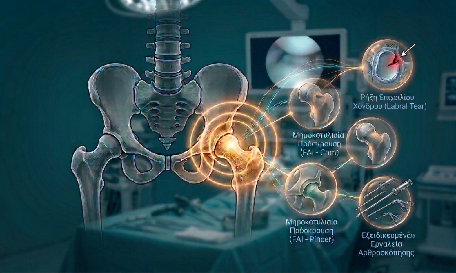
Hip Pain in Young and Active Patients: Pathologies, Symptoms, and Joint Preservation
Hip pain in young and active individuals often presents a diagnostic challenge. Symptoms frequently misdiagnosed as simple muscle strains, tendinitis, or lower back pain often originate from intra-articular hip pathology. Early diagnosis and a Joint Preservation strategy are critical, as chronic mechanical strain can lead to premature cartilage degeneration and early-onset osteoarthritis.
Classic Clinical Symptoms
Patients with structural hip deformities or intra-articular damage typically present with a specific symptom pattern:
-
Pain Localization (The "C-Sign"): Pain is rarely confined purely to the posterior gluteal region. Instead, it is felt deeply within the joint, radiating primarily to the groin, anterior thigh, or trochanteric area. Patients characteristically describe this location by cupping their hand in a "C" shape around the lateral hip between the thumb and index finger.
-
Mechanical Symptoms: Catching, clicking, locking sensations, or transient feelings of instability during movement.
-
Exacerbation with Activity: Pain intensifies following athletic exertion, as well as during positions that increase intra-articular pressure—such as deep squatting, prolonged sitting (e.g., while driving), or rotational torso movements.
Primary Causes of Hip Pain in Active Patients
-
1. Femoroacetabular Impingement (FAI)
FAI is the leading anatomical cause of hip pain in young adults. It results from abnormal osseous morphology that causes premature mechanical contact during joint motion:
-
Cam Morphology: Bony prominence at the femoral head-neck junction.
-
Pincer Morphology: Acetabular overcoverage of the femoral head.
-
Mixed Type: A combination of both Cam and Pincer deformities (most common clinical presentation).
-
-
2. Acetabular Labral Tear
The labrum is a fibrocartilaginous ring encompassing the acetabular rim that maintains joint stability and the crucial fluid seal (suction seal mechanism). Repetitive mechanical micro-trauma from FAI or acute injuries can tear or detach the labrum, causing sharp pain and mechanical joint symptoms.
Complex Structural Deformities: Dysplasia & Version Abnormalities
In select cases, hip pain stems not only from localized impingement, but from broader structural biomechanical alignment issues:
-
Hip Dysplasia: The acetabulum is shallower than normal, offering inadequate coverage of the femoral head. This results in joint instability, increased focal loads on the labrum, and accelerated joint wear.
-
Femoral / Acetabular Version Abnormalities: Horizontal alignment of the femur and acetabulum significantly impacts mechanics:
-
Excessive Anteversion: Anterior tilting of the femur or acetabulum alters gait mechanics.
-
Retroversion: Backward orientation of the acetabular rim or femoral neck, causing severe anterior structural impingement.
-
Diagnostic Note: When arthroscopic repair is performed in the presence of underlying severe dysplasia or major torsional abnormalities (anteversion/retroversion), isolated arthroscopy may be insufficient. Specialized cases may require corrective osteotomies (such as Periacetabular Osteotomy - PAO or Femoral Torsional Osteotomy) to re-align the skeleton and ensure long-term joint survival.
Modern Management: Hip Arthroscopy & Joint Preservation
Modern Orthopedics emphasizes native joint preservation through minimally invasive hip arthroscopy:
-
Osteoplasty: Surgical bony reshaping (Cam/Pincer resection) to restore normal clear space and eliminate impingement.
-
Labral Repair: Re-fixation of the torn labrum back to the acetabular rim using bioabsorbable anchors, successfully restoring the fluid suction seal.
-
Chondroplasty & Microfracture: Targeted management of concurrent articular cartilage defects to stimulate repair.
Persistent groin pain or mechanical clicking should not be managed solely with temporary pain relief. Specialized clinical evaluation and advanced imaging represent the essential first steps toward protecting joint longevity and restoring functional quality of life.


