
News and Announcements
Chondral Lesions & Subchondral Cysts in the Hip: Diagnosis & Arthroscopic Management
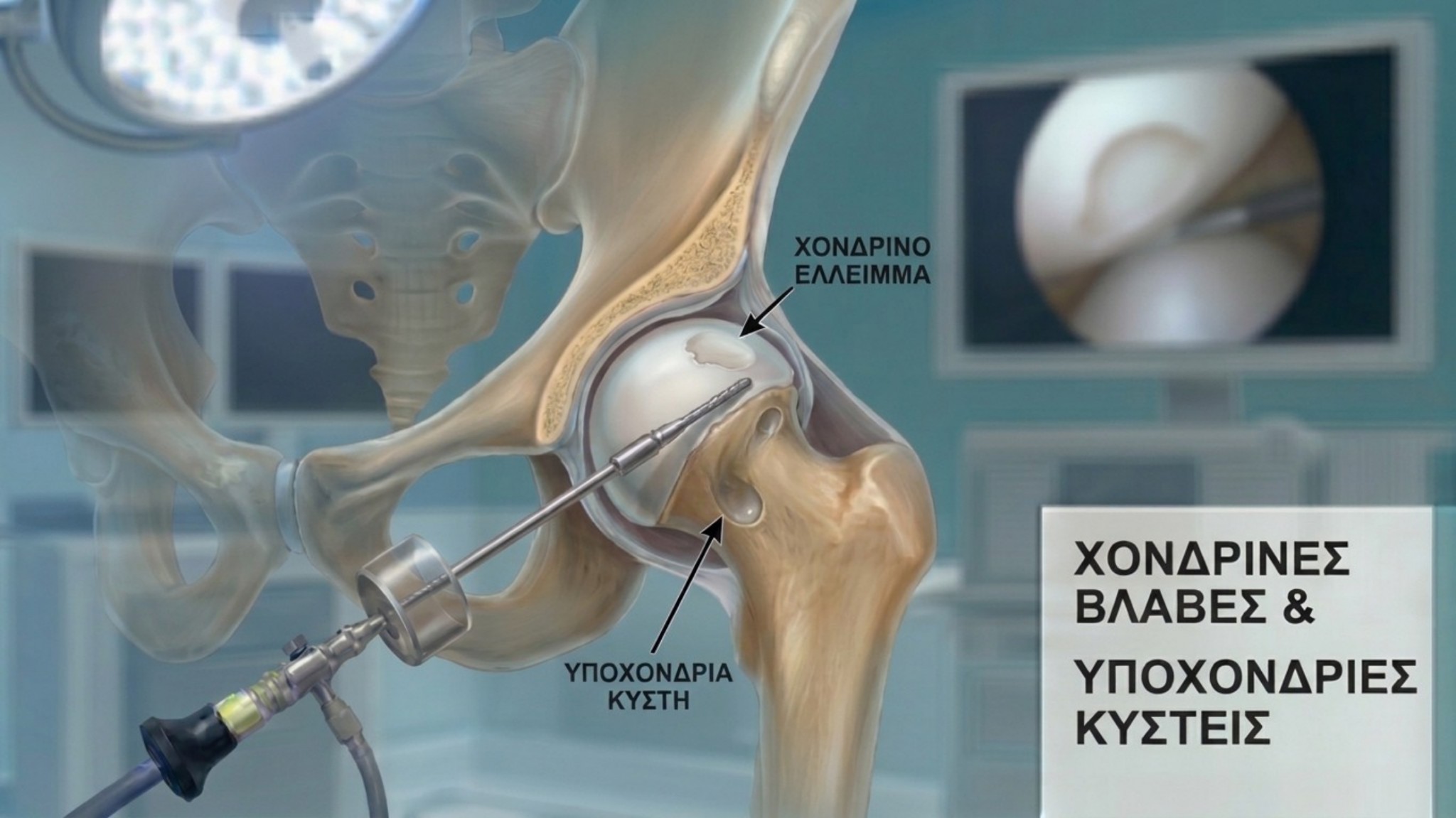
Modern Orthopaedics places a strong emphasis on preserving the natural hip joint (Joint Preservation). When a structural variance, such as Femoroacetabular Impingement (FAI) or a labral tear, remains unaddressed, the load distribution across the joint shifts. This alteration can gradually affect both the articular cartilage and the underlying bone.
Timely identification of these changes allows for effective joint protection, preserving the patient's quality of life and active lifestyle.
1. What are Chondral Lesions?
Articular cartilage is a highly smooth tissue covering the joint surfaces (the femoral head and acetabulum), ensuring frictionless gliding of the bones during movement.
When localized and repetitive mechanical pressure is applied — as seen in femoroacetabular impingement — the cartilage may experience focal stress. This manifests as:
-
Initial Surface Alteration: The early stage where mild changes in the consistency of the cartilage surface occur.
-
Focal Chondral Defect: A localized area of cartilage wear or detachment at a specific site within the joint.
Addressing these defects in their early stages is essential, as articular cartilage lacks its own blood supply to fully recover without targeted intervention.
2. How Do Subchondral Cysts Form?
The bone situated directly beneath the articular cartilage is called the subchondral bone. When the overlying cartilage develops a defect, mechanical loads are transferred more directly to the subchondral bone.
As a natural response to this increased pressure, the bone may develop bone marrow edema (focal stress) or subchondral cysts. These are small, benign cavities that form within the bone due to increased intra-articular pressure.
Key characteristics and symptoms include:
-
A deep sense of pressure or discomfort in the groin region, which may also occur during rest.
-
Increased sensitivity following prolonged activity or extended sitting.
3. Diagnostic Approach
Accurately mapping the extent and location of these changes is essential for designing an appropriate treatment plan.
-
Specialized Radiographic Imaging: Confirms the structural anatomy of the pelvis and femoral head.
-
Magnetic Resonance Imaging / Arthrography (MRI / MRA): The gold-standard investigation. It provides high-definition images, allowing for a detailed evaluation of cartilage thickness, structural integrity, and the presence of subchondral cysts or edema.
4. Arthroscopic Management & Restoration
Hip Arthroscopy is a modern, minimally invasive technique that allows the surgeon to correct the primary cause (such as resecting the FAI bone morphology) while simultaneously treating and restoring joint health in the same procedure.
Arthroscopic joint preservation and restorative techniques include:
-
Arthroscopic Chondroplasty: Precise smoothing of unstable cartilage margins to create a uniform surface and prevent further friction.
-
Bone Marrow Stimulation (Microfractures): Creating microscopic channels in the subchondral bone to stimulate the body's natural response for producing protective fibrocartilage.
-
Biological Therapies & Augmentation: Targeted application of biological agents (such as Platelet-Rich Plasma - PRP) to support tissue recovery.
-
Decompression of Subchondral Cysts: Debridement of the cysts and, when necessary, filling with biocompatible graft materials to support bone remodeling.
Conclusion
Chondral lesions and subchondral cysts of the hip are manageable conditions when diagnosed early. Thanks to advances in Hip Arthroscopy, we now possess the techniques to protect the natural joint, relieve symptoms, and support patients in maintaining their active lifestyle.


