News and Announcements
Femoroacetabular Impingement (FAI): Cam vs Pincer – The Primary Cause of Hip Pain in Active Young Adults
Hip pain in young and active adults is frequently misdiagnosed as a temporary muscle strain. However, modern Orthopaedics has demonstrated that the most common underlying cause of intra-articular lesions in the young hip is Femoroacetabular Impingement (FAI).
What is Femoroacetabular Impingement (FAI)?
In a normal hip joint, the spherical femoral head rotates smoothly within the acetabulum (the hip socket). In femoroacetabular impingement, an anatomical mismatch occurs due to bony overgrowth on either the femoral head or the acetabular rim (or both).
This structural abnormality prevents the full, unhindered range of motion of the joint, causing repetitive mechanical friction—particularly during hip flexion and internal rotation (e.g., deep squatting, driving, or athletic activities)—which leads to progressive wear and tear of the intra-articular structures.
The Three Morphological Types of Impingement: Cam, Pincer & Mixed
The pathology is classified into three main categories based on the anatomical location of the bony abnormality:
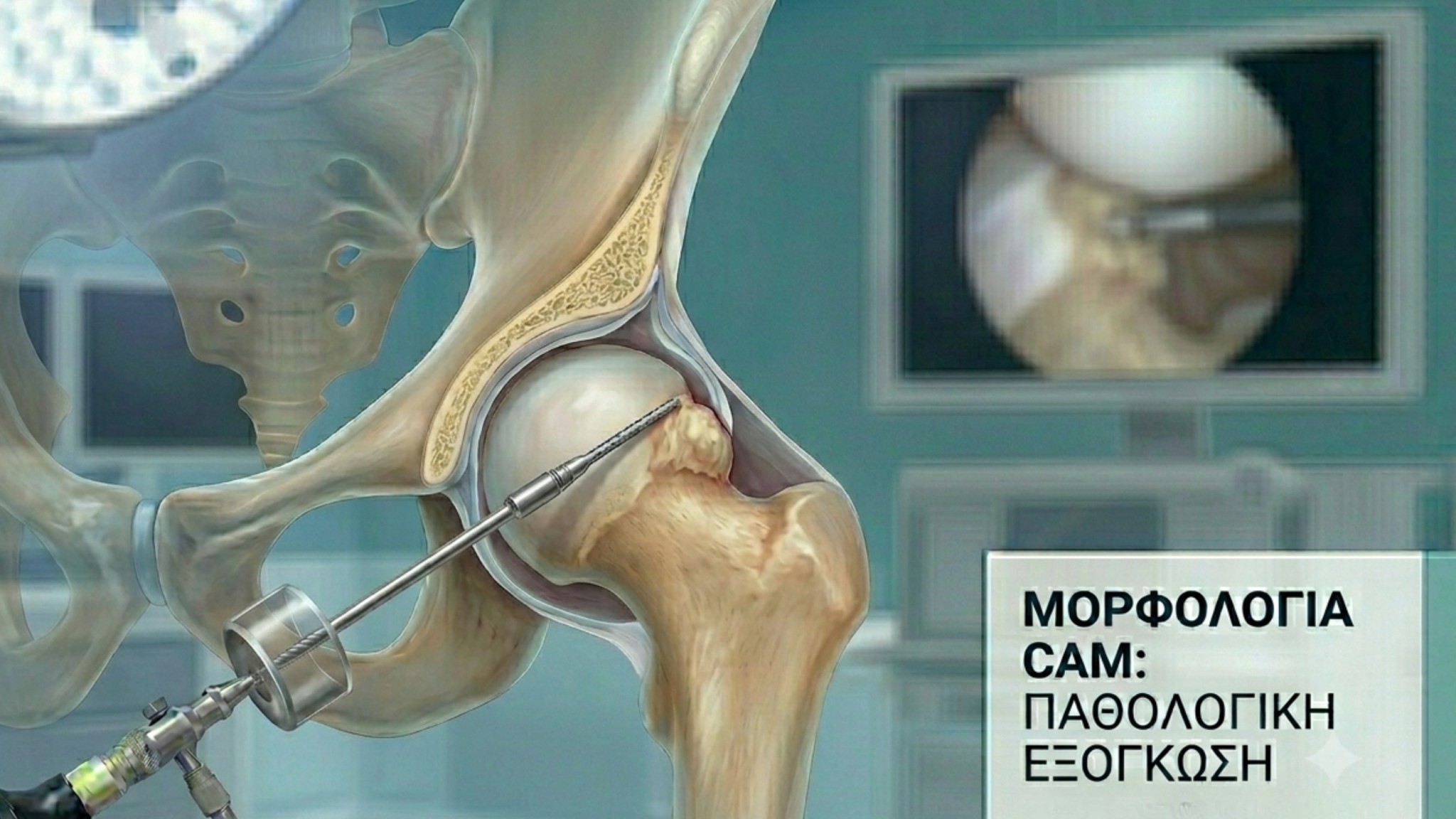
1. Cam-type Impingement
-
Anatomical Defect: The femoral head loses its perfectly spherical shape, presenting a bony prominence ("bump") at the femoral head-neck junction.
-
Mechanism of Injury: During hip flexion and internal rotation, this bony prominence impinges against the anterior acetabular rim, causing gradual degeneration, tearing, or avulsion of the articular cartilage and acetabular labrum.
-
Patient Profile: Most commonly observed in young males and high-impact athletes (football, track and field, CrossFit).
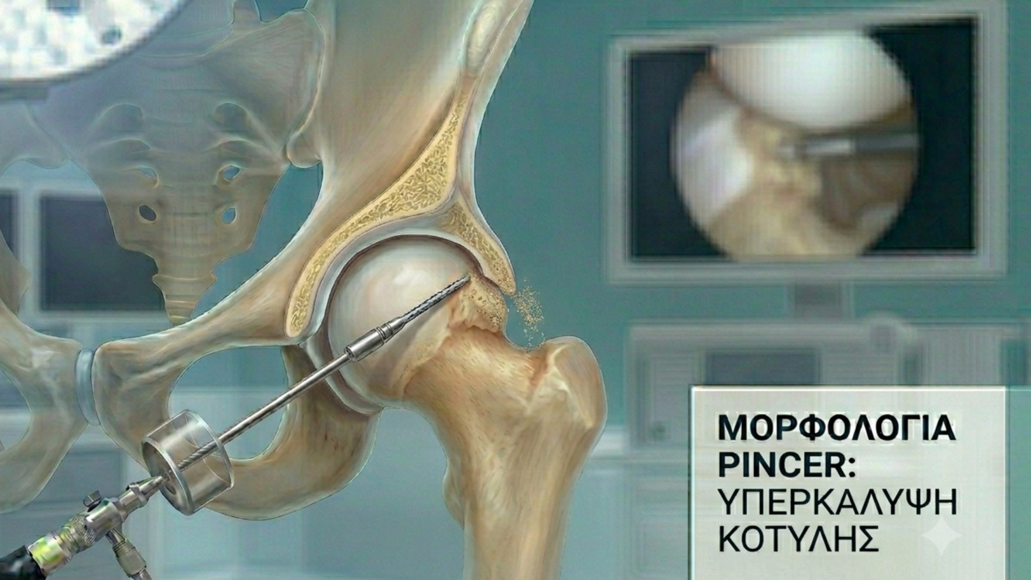
2. Pincer-type Impingement
-
Anatomical Defect: The acetabulum overcovers the femoral head, either because the socket is deeper than normal or because the anterior acetabular rim extends excessively.
-
Mechanism of Injury: The acetabular rim acts as a pincer, compressing and tearing the labrum against the femoral neck at the limits of joint motion.
-
Patient Profile: More frequently encountered in active, middle-aged females.
3. Mixed-type Impingement
This is the most common presentation in clinical practice (accounting for over 70% of cases), characterized by the simultaneous presence of both Cam and Pincer deformities.
Clinical Presentation: Symptoms & Signs
Femoroacetabular impingement develops gradually and presents with a distinct clinical pattern:
-
Location of Pain: Deep, dull, or sharp groin pain that may radiate to the anterior thigh or the trochanteric region (outer hip).
-
Mechanical Symptoms: Clicking, catching, locking sensations, or restricted range of motion.
-
Exacerbating Factors: Symptoms intensify after athletic activities or prolonged sitting (e.g., during long drives or desk work).
From Impingement to Labral Tears and Osteoarthritis
The acetabular labrum is a fibrocartilaginous ring surrounding the socket that maintains joint seal and stability.
Repetitive mechanical friction from Cam or Pincer impingement inevitably leads to labral tears and progressive destruction of the articular cartilage. If left untreated, this ongoing joint damage predictably accelerates the onset of early hip osteoarthritis.
Diagnostic Approach
Timely and accurate diagnosis is based on a comprehensive evaluation:
-
Specialized Clinical Examination: Impingement tests that reproduce the patient’s symptoms.
-
Specific Radiographic Views: Plain X-rays (e.g., AP pelvis, Dunn view) to identify Cam and Pincer bony deformities.
-
Magnetic Resonance Imaging / Arthrography (MRI/MRA): High-resolution imaging of choice to thoroughly assess the integrity of the labrum and articular cartilage.
Modern Treatment Options: Hip Arthroscopy
When conservative management (activity modification, physical therapy) fails to provide lasting relief, the definitive solution is Hip Arthroscopy.
This is a minimally invasive surgical procedure performed through small portals (2–3 mm), allowing for complete anatomical restoration:
-
Arthroscopic Osteoplasty: Precise reshaping and removal of the bony prominence (Cam/Pincer resection) to eliminate impingement permanently.
-
Labral Repair & Refixation: The torn labrum is anatomically reattached to the acetabular rim using bioabsorbable anchors, restoring the joint's suction seal.
Conclusion
Femoroacetabular impingement should not be dismissed as occasional muscle soreness. Early diagnosis and arthroscopic intervention prior to the onset of irreversible cartilage damage represent the cornerstone of Joint Preservation. This approach allows patients to preserve their native joint and safely return to their athletic activities.
Dr. Alexandros Tzaveas, specializing in advanced Hip Arthroscopy techniques, provides individualized diagnostic and surgical care in Athens and Thessaloniki.