
News and Announcements
Indications for Hip Arthroscopy: Which conditions are treated arthroscopically and when is the procedure selected?
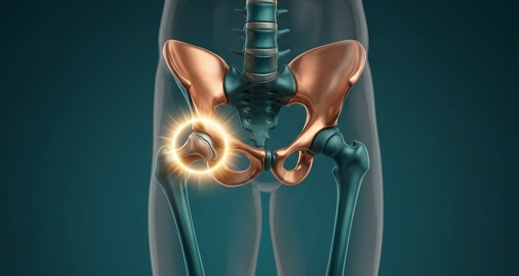
Hip Arthroscopy: Indications and Pathology Management
Hip arthroscopy represents one of the most advanced, minimally invasive procedures in modern Orthopedics. It allows the specialized surgeon to evaluate the interior of the joint with exceptional clarity through portal micro-incisions and perform complete arthroscopic repair of pathologies that previously required large, open surgical approaches.
Primary Indications for Hip Arthroscopy
-
1. Femoroacetabular Impingement (FAI – Cam & Pincer):
FAI is the primary anatomical cause of hip pain in young and active patients, stemming from abnormal osseous morphology:
-
Cam Type: A bony prominence (bump) located at the femoral head-neck junction.
-
Pincer Type: Excessive coverage of the femoral head due to overgrowth of the acetabular rim.
-
Mixed Type: The most common clinical presentation, combining both Cam and Pincer deformities.
-
Intervention: Precise arthroscopic osteoplasty and chondroplasty reshape the bone, eliminating mechanical impingement and restoring normal joint range of motion.
-
-
2. Acetabular Labral Tear:
The labrum is a fibrocartilaginous ring lining the acetabular rim that maintains joint stability and the crucial suction seal mechanism. FAI or acute trauma can cause labral detachment or tearing, leading to deep groin pain and mechanical symptoms (clicking, catching, or locking).
-
Intervention: Standard of care involves arthroscopic refixation and repair using bioabsorbable suture anchors to restore original anatomy, outperforming tissue resection (debridement) in preserving long-term joint integrity.
-
-
3. Early Osteoarthritis & Articular Cartilage Lesions:
In early-to-moderate cartilage degradation, surgical intervention focuses on Joint Preservation principles.
-
Intervention: Arthroscopic debridement removes loose chondral fragments and inflammatory tissue to relieve pain. Microfracture techniques (bone marrow stimulation) can be applied to encourage cartilage repair and delay or prevent total joint replacement.
-
-
4. Synovial Chondromatosis:
A benign condition where the synovial membrane undergoes metaplasia, producing multiple loose cartilaginous bodies within the joint space. These fragments cause severe pain, joint effusion (swelling), and locking.
-
Intervention: Complete arthroscopic extraction of loose bodies combined with targeted synovectomy minimizes post-operative pain and speeds recovery.
-
-
5. Paralabral Cysts:
Cysts that form secondary to labral tears when synovial fluid leaks through the defect into surrounding extra-articular tissues, often compressing adjacent nerves and causing severe radiating pain.
-
Intervention: Decompression and drainage of the cyst combined with arthroscopic repair of the underlying labral tear to eliminate recurrence.
-
-
6. Chronic Hip Synovitis:
Persistent inflammation of the synovial membrane caused by micro-trauma, overuse, or mechanical irritation, resulting in chronic deep pain, pressure, and fluid accumulation.
-
Intervention: Targeted subtotal or total synovectomy selectively resects inflamed tissue, reducing intra-articular pressure and preventing secondary cartilage degradation.
-
Hip arthroscopy provides an etiology-based treatment that corrects mechanical anatomical defects and protects the joint from premature wear. Modern Joint Preservation protocols—often delivered via One Day Clinic pathways—ensure targeted care aimed at a rapid, safe return to full functional mobility.


